Cores Resources
Center Cores are designed to consolidate and increase access to existing resources for vision research. Most of the core resources are located in the Vision Research Core in Biotech Place.
Visual Physiology and Behavior Core
Contact: Jay Ma, MD, PhD Jianxing.Ma@wfusm.edu
Purpose: Induction of Ocular Pathology and In Vivo Measurement of Visual and Retinal Function
Equipment:
Oxygen cycler
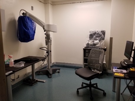
Laser Photocoagulation System
Optomotry System
Pelli Robson Eye Chart
Frisby Stereoacuity Plates
Trial lenses and trial frame
Electroretinogram – Ganzfeld Dome
Pattern Electroretinogram and Visual-evoked Potentials
Visual Imaging Core
Contact: Jay Ma, MD, PhD Jianxing.Ma@wfusm.edu
Purpose: Photography of the retina and eye
Equipment:
Optical Coherence Tomography-angiography
Optical Coherence Tomography
Fluorescent fundoscopy
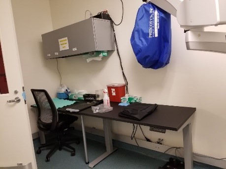
Precision Vision LED ETDRS Illuminator Cabinet
Traceable® Dual-Range Light Meter
Vision Metabolism and Cell Signaling Core
Contact: Rebecca Sappington, PhD rebecca.sappington@wfusm.edu
Purpose: Metabolic and cell signaling profiling
Equipment:
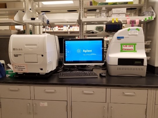
Seahorse Metabolic Profilers (x3)
Oroboros Metabolic Profilers (x4)
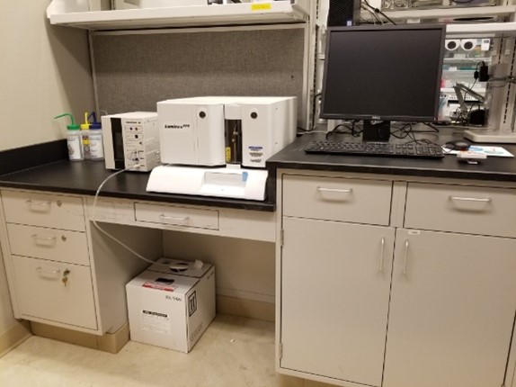
Luminex P200 Multiplex ELISA
Ocular Histopathology Core Resource
Director and Contact: Rebecca Sappington, PhD rebecca.sappington@wfusm.edu
The Ocular Histopathology Core Resource (OHCR) in the Translational Eye and Vision Research (TrEVR) Center seeks to accelerate discovery in the vision sciences by providing services and resources for ocular tissues and fluids.
Ocular Biorepository:
- Collection and curation of fixed ocular samples from non-vision studies, including rodent, swine and non-human primate
- Network of frozen and fixed samples from TrEVR Center members, including rodent models of eye disease and human donor eyes with diabetic retinopathy and age-related macular degeneration
Ocular Histopathology:
- OCT Embedding and Cryosectioning
- EPON Embedding and Semi-thin Sectioning
- Basic Histology
- Light and confocal microscopy
- In situ assays
- Special services at discretion of Director, i.e. training, tear film collection and sample preparation